Uppgift
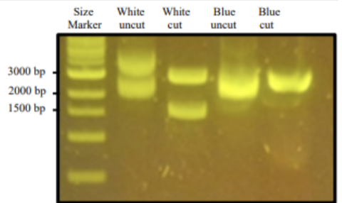
A gene of interest was amplified and verified using the blue/white screening method. The plasmids from two bacterial colonies, one blue colony and one white colony, were purified. Then restriction enzyme digestion was performed, and the samples were run on a gel as follows: white uncut, white cut, blue uncut, blue cut (see figure below). After imaging the gel, we can conclude that while there is plasmid DNA in all samples, the white sample was probably mixed with the blue sample. Explain what finding in the gel indicates the white sample was probably mixed with the blue sample by referring to the size/number of bands. Account for the other source of error for this outcome besides pipette tip contamination from sample to sample. (4p) (Max 200 words)
OBS! This question needs to be answered in English.
Answer
the findings in the gel that indicate that the samples were mixed are the lines in the white uncut sample. It has two visible bands, with one lining up quite nicely with the blue uncut bands in size. If the plasmid is uncut, than there shouldn't be two distinct bands because the sample only consists of an uncut product. The white uncut should also have a larger amount of base pairs than the blue uncut because we have transformed a gene of interest into it, but they seem to have the same amount.
a source of error for this outcome could be that the colony picked for the white cut may have been close to a blue colony - causing a small amount of the blue colony to be accidentaly picked.
Totalpoäng: 4