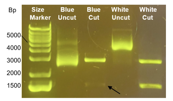
You amplified a gene of interest using the blue/white screening method. You extracted plasmids from two bacterial colonies (one blue colony and one white colony), then performed restriction enzyme digestion and ran the samples on a gel as follows: blue uncut, blue cut, white uncut, white cut (see gel below). When imaging the gel, however, you notice there is a faint band in the blue cut sample (see arrow), which should not be there. Explain why the faint band should not be present in that sample and what are the possible sources of error for this outcome? Refer to the size/number of bands in comparison to the bands in the other lanes for your explanation. This questions needs to be answered in English. (4p)
Answer
The blue samples are samples with no DNA-insert.
They have an intact lacZ-gene and are able to produce β-galactosidase which breaks down X-gal to a blue pigment.
Since the blue colony samples have no DNA insert, the blue sample should show one band on the gel electrophoresis.

The reason that the blue cut shows two bands can be because a mixed colony was chosen, meaning one that also had white colonies.
This could likely be the case because the second band has a 1500 bp size which the second band of the "white cut" also has.
Another possible reason for this odd result is contamination.
The "blue cut" sample could possibly have been contaminated with white cut samples when pipetting or by improper labelling of tubes.
This could be the case as the samples in both lanes show similar sizes (even the larger band is approximately the same size in both, 3000 bp).